What Is 3D Dental Imaging and How Does It Work?
3d dental imaging has changed how clinicians examine teeth, bone, nerves, and surrounding anatomy. Unlike a flat intraoral radiograph, cone-beam computed tomography creates a three-dimensional dataset. A rotating scanner captures many projections around the patient’s head. Software then reconstructs these views into cross-sectional images. Clinicians can inspect the jaw from axial, sagittal, and coronal directions.
The numbers explain its growing role. Fortune Business Insights estimates that the global dental imaging market will expand substantially through 2032. MarketsandMarkets also identifies cone-beam computed tomography as a major growth segment. However, market forecasts vary between reports. That difference deserves attention. Commercial growth does not automatically prove clinical necessity.
Oral and maxillofacial radiologist Dr. William C. Scarfe has emphasized, “CBCT should be considered when conventional radiography does not provide the information required.” This principle supports responsible imaging. The American Dental Association and U.S. Food and Drug Administration similarly recommend patient-specific justification and dose optimization. A scan should answer a clear clinical question.
Imagine a lower molar beside the mandibular nerve. A three-dimensional view may reveal their distance before implant placement or surgery. It can also expose hidden root anatomy, bone defects, or impacted teeth. Still, sharper images are not always better. Larger fields may increase radiation exposure and create incidental findings. Interpretation also requires training.
The technology is powerful, but not magical. Its value depends on careful selection, accurate positioning, suitable exposure settings, and qualified review. Patients deserve both useful images and honest explanations. When those safeguards are missing, 3d dental imaging can create confusion instead of clarity.
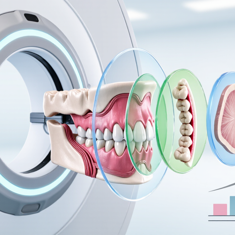
3D dental imaging is a method for viewing teeth, bone, and nearby structures in three dimensions. Unlike a flat dental X-ray, it can display a region from different angles and depths. A common method uses cone-beam computed tomography, often called CBCT. During the scan, a small X-ray unit rotates around the patient’s head. It captures multiple images within seconds. Computer software then combines them into a detailed 3D model made of tiny units called voxels. The image may reveal tooth roots, jawbone density, nerves, sinuses, and unerupted teeth. It is not a photograph. It is a calculated representation.
The patient usually sits or stands still and gently bites a support. Movement can blur important details. Dentists may request 3D imaging before implant placement, complex root canal treatment, orthodontic planning, or evaluation of impacted teeth. A qualified dental professional should compare the scan with symptoms, medical history, and a physical examination. One scan cannot explain every problem. It can also contain artifacts from metal fillings or dental movement. Exposure levels vary by equipment and scan size, so imaging should have a clear clinical reason. Pregnancy, previous radiation exposure, and other health details should be discussed beforehand.
A bright, convincing image may still require careful interpretation. That limitation deserves attention.
Three-dimensional dental images come from several technologies, each capturing different details. Cone beam computed tomography, or CBCT, uses a rotating X-ray beam. It creates many thin images around the patient’s head. Software then reconstructs them into a three-dimensional view of teeth, bone, nerves, and sinuses. The result resembles a small digital model that clinicians can rotate on a screen.
Optical intraoral scanners use a handheld camera and structured light. The device captures thousands of surface measurements across the teeth and gums. Software joins these measurements into a color 3D model. This method is comfortable and radiation-free. It works well for monitoring tooth movement, planning restorations, and checking how teeth meet. However, saliva, movement, and deep gum margins can reduce accuracy. It sounds simple. Not quite.
Some practices also use facial scanning or dental photogrammetry. These systems record facial shape, jaw position, or implant locations. Clinicians may combine this information with CBCT and surface scans. That combination can improve planning, but it demands careful alignment and trained judgment. A poorly calibrated scanner may produce a convincing image with small errors. I once assumed a detailed model was automatically reliable; clinical imaging requires more caution. Dentists should select the technology according to the diagnostic question, not visual appeal. CBCT also involves radiation, so professional guidelines recommend using it only when the expected benefit justifies the exposure.
A 3D dental scan uses cone-beam computed tomography to create detailed images of teeth, bone, nerves, and surrounding structures. Unlike a standard dental X-ray, it captures information from multiple angles. The result is a three-dimensional view that can support implant planning, orthodontic assessment, and complex diagnosis.
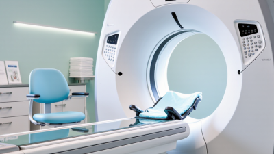
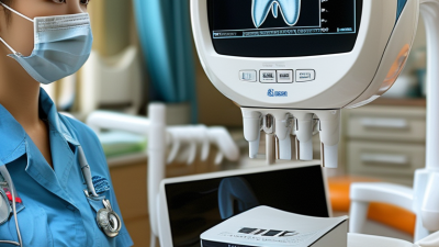
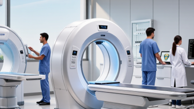
The appointment usually takes place in a dental imaging room. The patient removes metal items, such as earrings, glasses, or removable appliances. A technician positions the head carefully and places a small support near the chin or forehead. The patient bites gently on a disposable guide and stays still. Then, the scanner rotates around the head for several seconds.
It is quick and painless.
The scan does not require injections or special preparation in most cases. Patients may hear a soft mechanical sound during the rotation. Movement can blur the images, so another scan might occasionally be needed. That is not ideal, but it happens. The radiation dose varies by the machine and scan area, so the smallest useful field should be selected. Afterward, a qualified dental professional reviews the images alongside the patient’s symptoms and clinical examination. A scan alone cannot explain every problem, and its findings need careful interpretation.
Three-dimensional dental imaging, often created with cone-beam computed tomography, builds a layered view of the mouth. Instead of a flat X-ray, it shows teeth, bone, nerves, and nearby spaces from multiple angles. The scan is quick. A patient usually sits while the unit rotates around the head. The detector records many projections, and software reconstructs them into cross-sectional images.
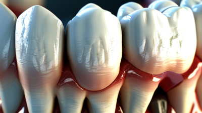
This view can reveal hidden root canals, curved roots, impacted teeth, and small fractures. It also maps jawbone height and width before implant planning. The lower jaw nerve canal may appear as a dark pathway near tooth roots. In the upper jaw, imaging can show sinus floors, cyst-like spaces, and inflammation-related changes. Orthodontic assessment may include tooth position, jaw growth, and crowding beneath the gumline.
However, a scan does not diagnose every problem by itself. Metal fillings can create streaks, while movement may blur important edges. Soft tissues are less clearly displayed than bone and teeth. A trained dental professional should compare the images with symptoms, examination findings, and standard radiographs. That interpretation matters. In practice, three-dimensional detail can clarify a difficult case, yet it may also invite overconfidence. The smallest visible irregularity is not automatically disease. Scan size and exposure should match the clinical question, especially for children and repeated examinations.
Dentists interpret 3D imaging as layered evidence, not a standalone answer. Cone-beam computed tomography creates cross-sectional slices from rotating X-ray measurements. Software reconstructs axial, coronal, sagittal, and curved views. The clinician checks each plane before reviewing the rendered volume. Patient history, examination findings, and earlier radiographs shape interpretation. A dark area may indicate infection, normal anatomy, or an artifact. Artifacts can mislead.
For implant planning, dentists measure bone height, width, and distance from the mandibular nerve or sinus. For impacted teeth, they trace root position and possible contact with neighboring roots. In endodontics, small-field scans may reveal missed canals, fractures, or lesions. Motion and metal can obscure details. The American Association of Endodontists and American Academy of Oral and Maxillofacial Radiology recommend CBCT only when it provides clinically useful information. Their position statement also supports the smallest suitable field of view.
Radiation dose remains part of responsible interpretation. FDA dental CBCT guidance reports approximate effective doses from 20 to 600 microsieverts, depending on equipment and settings. NCRP Report 177 and ICRP Publication 129 emphasize justification, optimization, and dose reduction. A dentist should document the reason for scanning and avoid casual repeat imaging. Complex findings may require an oral and maxillofacial radiologist’s review. A neat 3D model can still support a wrong assumption. Image quality, anatomy, and clinical uncertainty require discussion with the patient.
Typical effective radiation doses for common dental imaging examinations
Values show approximate representative effective doses in microsieverts (µSv); actual exposure varies with equipment, scan settings, field of view, and patient size. Intraoral, panoramic, and cephalometric images are mainly 2D, while cone-beam CT creates volumetric 3D data. Dentists interpret 3D images by reviewing cross-sectional slices and reconstructed views to assess bone levels, tooth roots, impacted teeth, airway structures, and implant-planning anatomy. They use the findings together with clinical examination and other diagnostic information.