A CBCT machine, or cone-beam computed tomography machine, creates detailed three-dimensional images of teeth, bones, nerves, and nearby tissues. Unlike a standard dental X-ray, it does not produce only a flat shadow. It rotates around the patient, capturing many projections from different angles. Software then reconstructs these projections into a three-dimensional view.
The process is precise, but not mysterious. A patient stands, sits, or lies still while the scanner completes one rotation. The X-ray source and detector move together. A cone-shaped beam passes through the head, while the detector records changing levels of absorption. The computer converts this information into cross-sectional slices. Clinicians can inspect the jaw from multiple directions, measure bone height, and trace the position of an impacted tooth.
Dr. Allan G. Farman, a respected oral and maxillofacial radiologist, wrote, “The use of CBCT in dentistry is increasing rapidly.” His observation remains relevant, although growth should not replace judgment. A CBCT machine can reveal useful anatomy, yet it cannot decide whether an examination is necessary. That decision belongs to a qualified professional, based on the patient’s symptoms, history, and clinical findings. The scan also has limits. Metal restorations may create bright streaks. Movement can blur delicate structures. Image quality varies between devices and settings. Small details may appear convincing but still require careful interpretation.
This article explains how a CBCT machine works, what its main components do, and why imaging settings matter. It also considers radiation exposure, diagnostic value, and practical limitations. The technology is powerful. It is not perfect. Understanding both sides supports safer, more informed imaging decisions.
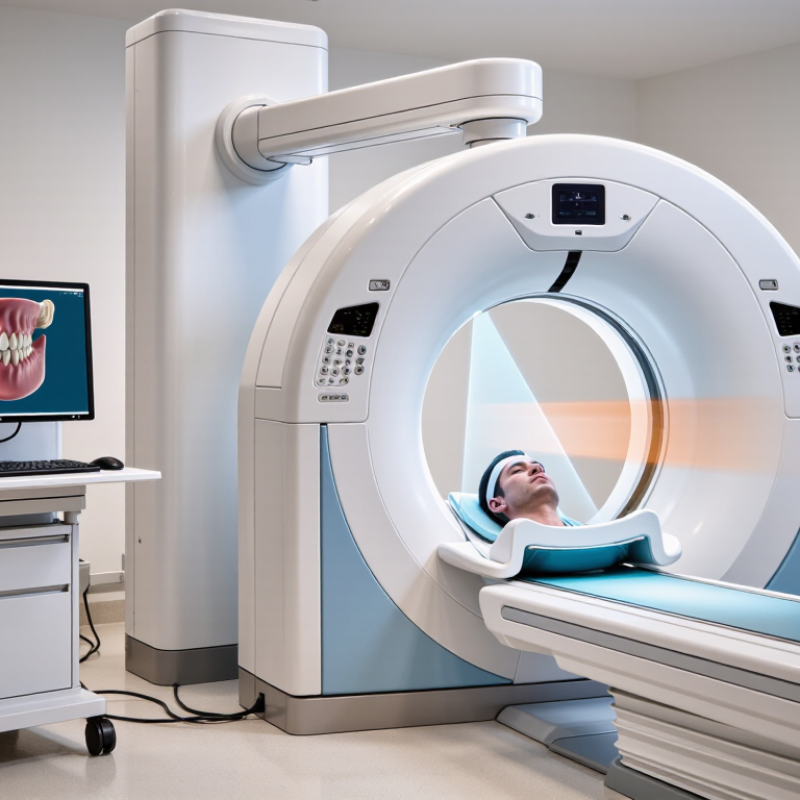
A CBCT machine, or cone-beam computed tomography system, creates three-dimensional images using a cone-shaped X-ray beam. The scanner rotates around the patient while a flat-panel detector captures many projection images. Reconstruction software then combines these views into small volumetric pixels, called voxels. The result shows teeth, bone, airways, and implant sites from multiple angles.
Its core purpose is targeted anatomical assessment. Dentists and radiologists may use CBCT to evaluate impacted teeth, jaw fractures, root anatomy, bone volume, or suspected lesions. A standard two-dimensional image can hide overlapping structures. CBCT reduces that limitation. It does not automatically provide a better answer, however. The scan must match a clear clinical question, and the field of view should remain as small as practical.
Radiation exposure varies considerably. The SEDENTEXCT project reported effective doses from approximately 11 to 1,073 microsieverts for dental CBCT examinations, depending on equipment and settings. The same report noted much lower typical doses for intraoral radiography. AAPM Report 175 also emphasizes patient-size selection, exposure control, and image-quality review. These figures are useful, but they are not universal. Protocols, anatomy, and reconstruction settings change the dose. A careful operator checks positioning, removes avoidable repeats, and records why three-dimensional imaging is needed. Small oversights matter.
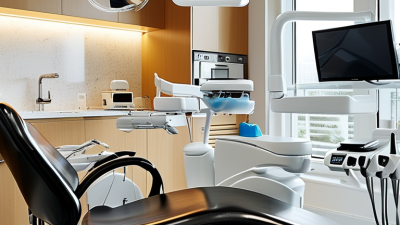
A cone beam computed tomography (CBCT) machine combines an X-ray source, detector, gantry, and computer system. Its main components work together to create detailed three-dimensional images. The X-ray tube produces a cone-shaped beam that rotates around the patient. A collimator narrows the beam and helps control the exposed area. This matters because unnecessary exposure should be minimized.
The flat-panel detector captures X-rays after they pass through the body. It converts those signals into digital data for processing. The gantry keeps the tube and detector aligned during rotation. A patient support holds the head or body in a stable position. Small details matter here. Even slight movement can blur delicate structures and reduce diagnostic confidence.
The control computer manages scanning settings, timing, and image storage. Reconstruction software turns hundreds of two-dimensional projections into cross-sectional and three-dimensional views. In clinical practice, trained operators adjust field size, resolution, and exposure according to the examination.
Higher resolution is not always better. It may increase radiation dose without adding useful information. Metal restorations can also create streaks that hide anatomy. No scan is perfect. Careful positioning, appropriate settings, and professional interpretation remain essential for reliable results.
A CBCT machine creates three-dimensional images by rotating an X-ray source and detector around the patient’s head. Unlike a conventional dental X-ray, it captures many projections from different angles. That rotation matters. The detector records the changing absorption of X-rays through bone, teeth, and soft tissue. Reconstruction software then combines these projections into small three-dimensional units called voxels. The finished volume can be viewed as cross-sectional slices, curved panoramas, or a rotating model.
During a scan, the patient usually sits or stands still while the system completes one rotation. Motion is the enemy. Even slight movement can create double edges or streaks around teeth and implants. Voxel size also affects detail. Smaller voxels may reveal a fine root fracture, but they can increase noise and radiation exposure. More detail is not automatically better. The U.S. Food and Drug Administration recommends using CBCT only when three-dimensional information can improve clinical decision-making.
Radiation dose varies widely with field of view, exposure settings, and scan duration. European Commission Radiation Protection No. 172 reports dental CBCT effective doses of approximately 11 to 1,073 microsieverts. That range is substantial. The International Atomic Energy Agency also emphasizes justification, patient positioning, and dose optimization. In practice, a focused scan may reduce unnecessary exposure while preserving useful anatomy. However, metal restorations can produce bright streaks that hide small structures. The image may look impressive, yet interpretation still requires trained professionals, clinical history, and careful review.
A cone-beam computed tomography (CBCT) machine rotates an X-ray source and a flat-panel detector around the patient. It collects many two-dimensional projection images from different angles, and computer reconstruction combines them into a three-dimensional volume made of small, usually isotropic voxels.
The chart shows representative voxel sizes commonly used for different CBCT applications. Smaller voxels provide finer spatial sampling, while the selected field of view, radiation dose, detector performance, and patient motion also affect the final image quality. Actual settings vary by clinical protocol.
A CBCT machine creates three-dimensional images from many X-ray projections. During a scan, the tube and detector rotate around the patient. The detector records changing patterns as X-rays pass through bone, teeth, and soft tissues. These measurements are not a finished picture. They are raw data.
Reconstruction software aligns the projections and calculates how much each tiny region absorbed. It converts those calculations into voxels, the three-dimensional equivalent of pixels. Thousands of voxels form cross-sectional views in multiple planes. They can also create a rendered volume for clinical inspection. The process is mathematical, not photographic. That distinction matters.
In practice, a qualified operator selects a suitable field of view and exposure setting. A small head movement can create blurred edges or double contours. Metal objects may produce bright streaks and dark gaps. Patient positioning also affects the final volume. It is easy to trust a sharp-looking image too quickly. I would treat it as measured evidence, not absolute reality. Image quality must be checked against the scan purpose and clinical findings. CBCT is valuable, but it does not replace professional interpretation or careful patient selection.
Cone-beam computed tomography (CBCT) creates detailed, three-dimensional images of bone and teeth. A cone-shaped X-ray beam rotates around the patient’s head or body. Sensors capture multiple views during one scan. Software then reconstructs them into cross-sectional and 3D images. CBCT is often faster than conventional medical CT. However, radiation exposure varies by scan size, settings, and patient needs. It does not replace a physical examination.
In dentistry, CBCT supports implant planning by showing bone height, bone width, and nearby nerve canals. Dentists may also use it to assess impacted wisdom teeth, complex root canals, jaw cysts, and facial trauma. Orthodontic teams can evaluate buried teeth and jaw relationships more clearly. For example, a scan may reveal whether an upper tooth lies close to the sinus floor. That detail can change the surgical approach. Still, every scan should answer a specific clinical question.
Medical applications commonly include sinus evaluation, temporal bone imaging, facial bone assessment, and selected extremity studies. CBCT can show fine bone structures, but it is less effective for many soft-tissue problems. Patient movement may create blurred edges or misleading lines. That limitation is easy to underestimate. Clinicians should compare CBCT findings with symptoms, examination results, and earlier images. A qualified professional must also review the full scan, not only the area that first attracts attention.
| Data Dimension | Typical Information | How It Works or Why It Matters |
|---|---|---|
| Imaging Modality | Cone-beam computed tomography (CBCT) | CBCT uses a cone-shaped X-ray beam and a digital detector to create three-dimensional images of anatomical structures. |
| Main Components | X-ray tube, flat-panel detector, rotating gantry, patient support, computer, and reconstruction software | The tube and detector rotate around the patient while the system collects multiple projection images for reconstruction. |
| Image Formation | Hundreds of two-dimensional projections are combined into a three-dimensional volume | Computer algorithms calculate the X-ray attenuation of tissues and produce axial, coronal, sagittal, and multiplanar views. |
| Field of View (FOV) | Commonly ranges from approximately 4 cm for a localized scan to more than 15 cm for larger anatomical coverage | A smaller FOV can focus on a specific region and may reduce radiation exposure; a larger FOV includes more anatomy but may increase dose. |
| Voxel Size | Often about 0.08–0.4 mm in dental and maxillofacial imaging | Smaller voxels can show fine structures more clearly, but they may require higher exposure and can produce more image noise. |
| Typical Scan Time | Usually about 5–40 seconds, depending on the examination protocol | Short acquisition times help limit motion, although patient movement can still create artifacts. |
| Radiation Exposure | Highly variable; commonly measured in the tens to several hundreds of microsieverts for dental examinations | Dose depends on FOV, tube current, tube voltage, voxel size, scan time, and the selected clinical protocol. The lowest dose that provides adequate diagnostic quality should be used. |
| Dental Implant Planning | Assessment of bone height, width, angulation, and proximity to vital structures | Three-dimensional visualization supports implant positioning and evaluation of the mandibular canal, maxillary sinus, and adjacent roots. |
| Endodontics | Evaluation of complex root anatomy, missed canals, fractures, resorption, and periapical lesions | Limited-FOV scans can provide detailed views when conventional two-dimensional radiographs do not answer a specific diagnostic question. |
| Orthodontics | Assessment of impacted teeth, skeletal relationships, airway anatomy, and craniofacial structures | CBCT may be useful when three-dimensional information is expected to affect diagnosis or treatment planning. |
| Oral and Maxillofacial Surgery | Evaluation of jaw lesions, facial trauma, impacted teeth, and surgical anatomy | The volumetric dataset helps identify the position and extent of bony structures before selected surgical procedures. |
| Ear, Nose, and Throat Applications | Selected evaluation of the paranasal sinuses, temporal bones, and certain airway structures | CBCT can provide high-resolution bone detail, although the appropriate modality depends on the clinical question and soft-tissue requirements. |
| Key Strengths | Three-dimensional imaging, high spatial resolution, relatively compact equipment, and fast acquisition | CBCT is particularly effective for teeth and bones, where fine structural detail is important. |
| Important Limitations | Limited soft-tissue contrast, possible metal and motion artifacts, and exposure to ionizing radiation | CBCT should be prescribed only when clinically justified, with the smallest suitable FOV and an optimized exposure protocol. |
Note: Technical specifications, radiation dose, scan time, and image quality vary by equipment, patient size, and examination protocol. Clinical use should follow professional guidelines and local regulations.