10 Essential Tips for Using Handheld Dental X Ray Effectively?
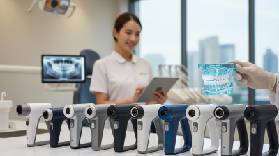
The use of handheld dental X-rays has revolutionized dental practices, enhancing diagnostic capabilities and patient care. According to a report from the American Dental Association, integrating portable imaging tools can reduce patient exposure to radiation by 50% compared to traditional methods. The convenience of handheld dental X-ray devices provides dental professionals with quick and accurate imaging results, which is crucial for timely treatments.
However, mastering the use of these devices is essential for achieving optimal results. Despite their advantages, professionals often face challenges, such as positioning and technique variations. A study published in the Journal of the American Dental Association found that improper technique can lead to misdiagnosis and increased need for retakes, highlighting the necessity of targeted training. By understanding both the strengths and limitations of handheld dental X-ray equipment, dentists can refine their approach and elevate patient outcomes.
Understanding the Advantages of Handheld Dental X-Rays in Practice
Handheld dental X-rays are transforming dental practices. They offer unique advantages that can enhance patient experience and improve diagnostic accuracy. One significant benefit is mobility. These devices allow dentists to take X-rays right at the chairside. This can reduce the time it takes to capture images. Patients stay comfortable and engaged when they don't have to move to a separate room.
Another key advantage is the reduced radiation exposure. Handheld X-ray units are designed to minimize radiation. Dentists can take precise images with lower doses. However, it is essential to calibrate and maintain these devices regularly. Proper training is also crucial to ensure effective use. Staff should understand the equipment’s limitations and challenges. This awareness helps minimize errors.
The compact design of handheld X-rays can sometimes lead to compromises in image quality. While they’re convenient, ensuring clarity in images is vital for an accurate diagnosis. Regularly reviewing the results and seeking opportunities for improvement is essential. Emphasizing clinical judgment alongside technology is crucial in enhancing patient outcomes. Balancing convenience with quality remains a challenge worth addressing in practice.
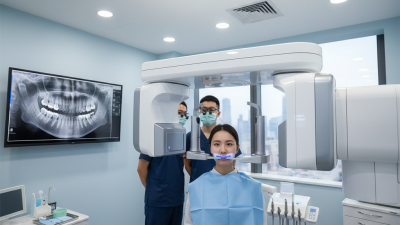
Key Safety Protocols for Minimizing Radiation Exposure in Dental X-Rays
Radiation exposure is a critical concern in dental practices. According to the American Dental Association, dental X-rays account for approximately 75% of the radiation exposure a person receives from medical sources. To effectively minimize this exposure, adhering to key safety protocols is essential.
Lead aprons and thyroid collars should be standard equipment for every patient receiving X-rays. Studies indicate that these simple tools reduce scattered radiation significantly. Another crucial protocol is to use the shortest exposure time possible, as this directly correlates with the amount of radiation received. The use of digital X-ray systems can further reduce radiation levels by 50% compared to traditional film methods.
However, despite advancements, there are still challenges in standardizing safety practices across all dental offices. Some practitioners may overlook key safety measures. Continuing education in radiation safety is vital. Promoting a culture of safety can ensure that both patients and staff are protected from unnecessary exposure while receiving essential diagnostic care.
Radiation Exposure Levels in Dental X-Rays
Best Practices for Positioning Patients to Optimize X-Ray Quality
Positioning patients correctly is vital for optimizing handheld dental X-ray quality. Research indicates that improper positioning can lead to poor image clarity, which may impede diagnosis. According to a study published in the Journal of Dental Research, up to 30% of X-rays taken may require retakes due to positioning errors. Ensuring that patients are comfortable allows for steadier images. Thus, clear communication about how they should position their head is essential.
For intraoral X-rays, angling the film parallel to the dental arch is crucial. The beam should be directed perpendicular to the film. This alignment minimizes distortion and enhances image details. Patients may need guidance on biting down or holding the film firmly. Additionally, using positioning devices can improve accuracy. A survey in Dental Clinics emphasized that correct usage of these devices decreased the error rate by 25%.
However, achieving optimal positioning can be challenging. Variations in patient anatomy and comfort levels often complicate this task. Practitioners must be adaptable. Continuous education and hands-on training are imperative in refining these techniques. Regular feedback sessions among dental staff can help identify common pitfalls. Embracing mistakes as learning opportunities fosters an environment of improvement and heightens patient care.
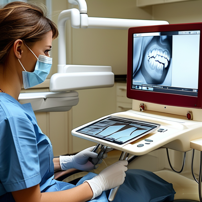
Interpretation of Handheld Dental X-Ray Images for Accurate Diagnoses
Interpreting handheld dental X-ray images accurately is crucial for effective diagnoses. Studies, such as one published in the Journal of Dental Research, show that 30% of dental issues can be missed due to poor image analysis. This highlights the need for a thorough understanding of the images produced by handheld devices.
Dental professionals must be aware of the positioning of patients and the angle of the X-ray beam. An improper angle may lead to distorted images. If the X-ray doesn't capture the area of concern correctly, minute details can be overlooked. Misinterpretation can have severe consequences, leading to improper treatment plans.
It's also important to recognize that even small adjustments in exposure time can drastically affect image clarity. The American Dental Association emphasizes consistent technique to enhance reliability. Analyzing images in a quiet room with adequate lighting can help practitioners focus better. Reflection on patterns and anomalies in the images can yield more accurate diagnoses.
Engaging in continual education and peer reviews can elevate interpretation skills over time.
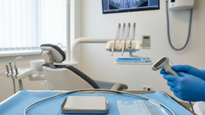
Maintenance and Calibration Tips for Handheld Dental X-Ray Devices
Proper maintenance and calibration of handheld dental X-ray devices are crucial for optimal performance. Regular checks ensure image quality and patient safety. Research shows that poorly maintained equipment can lead to inaccurate diagnoses. Clinicians should aim for precision to foster trust among patients.
One essential tip is to clean the device frequently. Wipe down the surface to prevent dust and debris accumulation. Use approved cleaning solutions. This step enhances the longevity of the equipment. Moreover, calibration should occur every six months, or as regulations suggest. Regular calibration confirms accurate radiation doses.
Finally, keep up to date with industry standards. Guidelines from organizations, like the American Dental Association, recommend periodic training on using these devices. Education prevents misuse and promotes safety. When in doubt, consult the user manual for specific maintenance instructions. By prioritizing these practices, dental professionals can ensure the reliability of handheld X-ray devices and enhance patient care.
10 Essential Tips for Using Handheld Dental X Ray Effectively
| Tip Number |
Tip Description |
Maintenance Activity |
Calibration Frequency |
| 1 |
Ensure Proper Battery Life |
Check battery levels before each use |
Monthly |
| 2 |
Use Protective Gear |
Inspect and replace gear as needed |
As required |
| 3 |
Clean the Device Regularly |
Wipe down with approved disinfectants |
After each use |
| 4 |
Check Image Quality |
Review and adjust settings for clarity |
Weekly |
| 5 |
Update Software Regularly |
Download and install updates |
Monthly |
| 6 |
Verify Radiation Exposure Settings |
Regularly calibrate exposure settings |
Bi-monthly |
| 7 |
Train Staff Properly |
Conduct regular training sessions |
Quarterly |
| 8 |
Store in a Safe Location |
Secure storage when not in use |
Always |
| 9 |
Use Correct Positioning |
Review positioning techniques regularly |
After each training |
| 10 |
Seek Professional Repairs |
Contact a technician for faults |
As needed |