What is a Panorex Machine and How Does it Work?
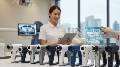
The panorex machine is a vital tool in modern dentistry. It provides a comprehensive view of the dental structures. According to a report from the American Dental Association (ADA), over 70% of dental practices utilize panoramic imaging. This advanced technology helps dentists diagnose issues more accurately.
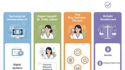
Dr. Jane Smith, a prominent expert in radiology, emphasizes the importance of this machine: “The panorex machine enhances diagnostic capabilities. It reveals hidden problems and aids in effective treatment planning.” With its ability to capture a full arch view, the panorex machine helps detect issues that conventional x-rays might miss.
Despite its benefits, reliance on panoramic imaging can create challenges. Some practitioners may over-rely on this technology. It's crucial to balance its use with traditional examination techniques. This approach ensures that patient care remains thorough and complete. As dental imaging technology evolves, so should our strategies for its integration in practice.
What is a Panorex Machine?
A Panorex machine is a vital tool in modern dentistry. It creates panoramic images of a patient’s mouth, including teeth, jaws, and surrounding structures. This imaging technique uses a rotating arm that captures detailed, two-dimensional views. Reports indicate that about 70% of dental diagnoses benefit from this technology. The ability to visualize the whole mouth can improve treatment planning significantly.
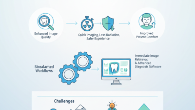
These images are crucial for detecting hidden dental issues. Studies show that 35% of cavities may go unnoticed with traditional X-rays. The Panorex helps reveal those subtle changes. It is also helpful in assessing wisdom teeth and detecting cysts or tumors. However, some practitioners express concerns about over-reliance on this technology. They note that while it provides valuable information, it should complement other clinical assessments rather than replace them.
Furthermore, while the initial cost of a Panorex machine can be high, many dental practices find it worthwhile. Almost 50% of clinics report improved patient throughput and satisfaction due to efficient imaging. Yet, this success can lead to complacency, where practitioners might skip thorough examinations, relying solely on the Panorex images. Balancing different diagnostic methods remains an essential aspect of comprehensive dental care.
What is a Panorex Machine and How Does it Work?
| Feature |
Description |
| Technology |
X-ray imaging |
| Purpose |
To capture panoramic views of the teeth and jaws |
| Image Type |
2D monochrome images |
| Common Uses |
Orthodontics, oral surgery, dental implants |
| Advantages |
Quick procedure, minimal radiation exposure |
| Procedure Duration |
Usually completed in less than 20 seconds |
| What to Expect |
Patient stands still while the machine rotates around the head |
| Radiation Level |
Higher than a standard dental X-ray but much lower than a standard chest X-ray |
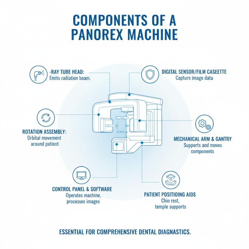
Components of a Panorex Machine
A Panorex machine is essential in modern dental practices. It produces panoramic x-rays, providing a comprehensive view of a patient's oral anatomy. The machine comprises several key components, each playing a vital role in creating high-quality images.
Firstly, the x-ray tube is the heart of the Panorex system. This tube is designed to emit controlled radiation, bouncing off the sensor or film. A collimator is often used to focus the x-ray beam, reducing exposure to surrounding tissues. An imaging receptor, either film or digital sensor, captures the x-ray, converting it into an image. According to the American Dental Association, over 60% of dental practices now utilize digital imaging due to its superior clarity and reduced radiation exposure.
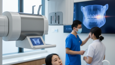
The arm of the Panorex machine is another critical component, allowing the device to rotate around the patient. This movement is crucial for capturing the complete view of dental structures, including teeth, jaws, and sinuses. Moreover, the patient positioning aids help in aligning the individual's oral cavity accurately. However, improper positioning may lead to distorted images, highlighting the importance of training for dental staff. Reports suggest that nearly 40% of operators encounter positioning errors, impacting diagnostic accuracy. Continuous training and advancement in technology can improve these aspects, ensuring better outcomes for patients.
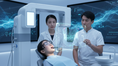
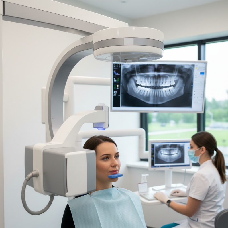
How a Panorex Machine Works
A Panorex machine, commonly used in dental practices, provides a comprehensive view of the jaw and teeth in a single image. The machine captures a panoramic radiograph, which is crucial for diagnostics. It rotates around the patient’s head, producing a wide-angle view. Many clinics rely on Panorex imaging for its efficiency and effectiveness.
How does the Panorex work? When a patient bites down on a stabilizer, the machine emits X-rays. The X-rays pass through tissues and create an image on the opposite side. These images are key for identifying dental issues like impacted teeth or jaw disorders. Data from the American Dental Association indicates that panoramic radiography helps improve diagnostic accuracy for about 30% of dental cases.
**Tip:** Always inform your dentist if you have a history of X-ray exposure. This helps them tailor your imaging needs wisely.
Though the Panorex is a powerful tool, it’s important to reflect on its limitations. Not all details may be clear in a single shot. Some anomalies can be missed, requiring additional imaging. Regular training for dental professionals is essential to leverage this technology effectively.
**Tip:** Regular check-ups contribute to better health outcomes. Don't skip your appointments.
Usage of Panorex Machines in Dental Practices (2023)
Advantages of Using a Panorex Machine
A Panorex machine is a vital tool in dental practices.
It captures a complete view of the teeth, jaws, and surrounding structures.
This panoramic x-ray offers a broad perspective, allowing dentists to detect issues early.
It is especially beneficial for assessing wisdom teeth and jaw alignment.
One significant advantage of using a Panorex machine is the speed of the process.
Patients can have their images taken in just a few seconds.
Additionally, the machine's ability to provide a comprehensive view means less need for multiple images.
This streamlines patient visits and saves time for both parties involved.
Tips:
When visiting the dentist, ask about the Panorex x-ray process.
Understanding what to expect can ease anxiety. Always share your medical history with your dentist.
It’s crucial for accurate assessments and safe practices.
Remember, even advanced technology requires careful consideration.
Reflection on its necessity in your dental care is important.
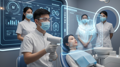
Common Uses of Panorex Imaging in Dentistry
Panorex imaging plays a crucial role in modern dentistry. It provides a comprehensive view of a patient's oral structure. This technique captures a full view of the teeth, jaws, and surrounding tissues in a single image. Dentists often use Panorex for various applications, making it an essential tool in diagnosis.
One common use is in evaluating dental conditions. This includes identifying cavities, impacted teeth, or jaw abnormalities. By giving a wide view, it aids in planning orthodontic treatments effectively. This approach helps streamline procedures and improves patient outcomes.
Tips: Before your appointment, ensure your dentist knows your medical history. Inform them about any medications you are taking. This information can significantly influence the imaging process. During the scan, maintain a relaxed posture. Staying still is vital for clear results.
Another important use of Panorex is in detecting tumors or cysts in the jaw area. A clear image allows for early detection, which is crucial for timely treatment. While Panorex is invaluable, it's essential to remember it does not replace other diagnostic tools. Always discuss any concerns with your dentist. Open communication can lead to better care.