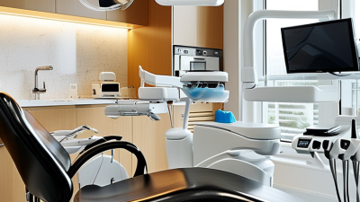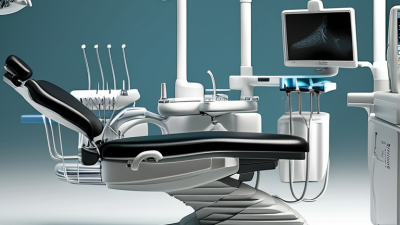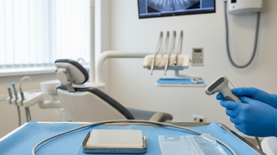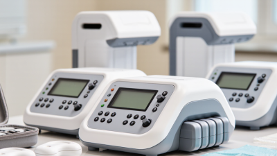
Oral disease affects nearly 3.5 billion people worldwide, according to the World Health Organization’s Global Oral Health Status Report 2022. That burden reaches ordinary clinics, where dentists must assess pain, missing teeth, bone levels, and developing dentition efficiently. A panoramic x ray machine can capture both jaws, the temporomandibular joints, and surrounding structures in one broad image. It offers a practical view before treatment begins.
The value is visible at the chairside. A patient sits upright, rests the chin, and keeps still for several seconds. The resulting image may reveal an impacted wisdom tooth, an unexpected lesion, or bone loss that a small intraoral image cannot show alone. The International Atomic Energy Agency and European radiological guidance stress justification, optimization, and quality assurance for dental imaging. These principles make equipment selection more important than impressive specifications.
A panoramic system can support orthodontic screening, implant assessment, oral surgery referrals, and routine documentation. It may also reduce repeated appointments when imaging is appropriately prescribed. Still, it is not a magic shortcut. Image distortion, patient movement, metal artefacts, and limited fine detail can affect interpretation. A panoramic image should not replace targeted intraoral views or three-dimensional imaging when clinical questions demand them.
The American Dental Association recommends selecting radiographs according to individual patient needs, not routine habit. That reminder deserves attention. Clinics should compare dose information, detector performance, positioning aids, software integration, maintenance support, and staff training. The best panoramic x ray machine is not simply the newest model. It is the system that produces clinically useful images, supports safe workflows, and fits the team’s real experience.
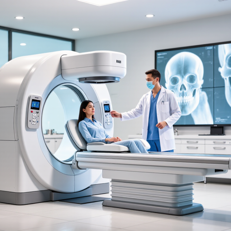
A panoramic X-ray machine creates a single, curved image of the mouth and jaws. The patient stands or sits while a rotating arm moves around the head. A chin rest and bite guide help keep the skull steady. The detector records X-rays from several angles. Software then combines these signals into one broad view. This image shows teeth, jawbones, joints, sinuses, and surrounding structures. It differs from intraoral images, which capture smaller areas with finer detail.
Clinics often use panoramic imaging for treatment planning, impacted teeth, orthodontic assessment, and jaw injuries. It can also reveal bone changes that are not obvious during a visual examination. The scan is quick, usually taking less than a minute after positioning. Patients should remain still and follow breathing instructions. Even a slight head tilt can create distortion. That matters. A trained operator checks positioning, exposure settings, and image quality before a dentist interprets the result. Professional judgment remains essential because panoramic imaging cannot replace every examination or targeted scan.
In practice, metal earrings, glasses, or removable appliances may produce bright artifacts. Removing them improves clarity and helps prevent unnecessary retakes. Radiation exposure is low, but clinics should follow local safety rules and use shielding when appropriate. Pregnancy concerns should be discussed before imaging, even when the examination seems routine. The machine is useful, not magical. Its broad view may blur fine root details or hide small cavities. The image should remain one carefully checked piece of evidence, not the whole diagnosis.
| Evaluation Dimension | Panoramic X-Ray Machine | Clinical Value for a Clinic |
|---|---|---|
| Imaging Modality | Extraoral two-dimensional dental imaging that captures a broad view of the jaws and facial structures. | Provides an overview before treatment planning without requiring an individual image for every tooth. |
| Typical Anatomical Coverage | Both dental arches, the mandible, portions of the maxilla, and selected surrounding structures. Depending on the field of view and positioning, the temporomandibular joints and maxillary sinuses may also be included. | Useful for screening, referrals, orthodontic assessment, oral surgery planning, and evaluating unerupted or impacted teeth. |
| Image Acquisition Method | The X-ray tube and image receptor rotate around the patient while the image is formed through a curved focal trough. | Creates a wide dental image in a single examination while helping reduce the need for multiple separate intraoral exposures. |
| Typical Acquisition Time | Commonly about 10–20 seconds for image capture, depending on the examination protocol and equipment configuration. | Supports efficient patient flow and can be practical for routine examinations and busy dental schedules. |
| Patient Positioning | The patient is usually standing or seated, with head supports and a bite guide used to align the teeth and jaws. | Allows imaging for patients who may find intraoral sensor placement uncomfortable or difficult. |
| Patient Comfort | The image receptor remains outside the mouth during exposure; the patient generally bites lightly on a positioning guide. | Can improve tolerance for children, patients with a strong gag reflex, limited mouth opening, or oral sensitivity. |
| Radiation Consideration | Radiation exposure is protocol-dependent and is generally lower than many cone-beam computed tomography examinations, although exact dose varies by equipment and settings. | Offers broad two-dimensional information when three-dimensional imaging is not clinically necessary. The examination should still be justified and optimized according to radiation-protection principles. |
| Common Clinical Applications | Assessment of tooth development, missing or impacted teeth, jaw fractures, advanced periodontal bone loss, extensive lesions, and general dental conditions. | Supports initial diagnosis, treatment planning, patient education, and referral decisions across several dental specialties. |
| Orthodontic Use | Shows the number, position, eruption path, and approximate root development of many teeth in one image. | Helps identify eruption problems, retained primary teeth, missing teeth, and possible root or bone-related concerns. |
| Oral Surgery Use | Provides a broad view of the jaws and can show the approximate relationship of impacted teeth to surrounding structures. | Useful for preliminary evaluation of third molars, retained teeth, fractures, and other conditions before deciding whether additional imaging is needed. |
| Image Characteristics | Produces a curved panoramic image with magnification and geometric distortion that can vary across the image. | Excellent for overview assessment, but precise measurements and detailed three-dimensional relationships may require another imaging method. |
| Comparison with Intraoral X-Ray | Covers a much larger anatomical area, while intraoral imaging generally provides higher detail for individual teeth and nearby supporting tissues. | Panoramic imaging complements rather than fully replaces bitewing, periapical, or occlusal examinations. |
| Comparison with CBCT | Provides a two-dimensional overview rather than volumetric three-dimensional data. | Panoramic imaging may be appropriate for broad screening, while CBCT is considered when three-dimensional information is clinically required. |
| Key Advantages | Wide anatomical coverage, relatively fast acquisition, extraoral positioning, and convenient examination workflow. | Can improve diagnostic efficiency and provide a useful first-line image for many routine and specialist dental assessments. |
| Important Limitations | Overlapping structures, image distortion, positioning errors, motion artifacts, and lower fine-detail resolution than many intraoral images. | Correct patient preparation, positioning, exposure selection, and clinical interpretation remain essential for reliable results. |
| Best-Practice Requirement | Use only when clinically justified, follow applicable radiation-safety regulations, and apply the lowest exposure that produces a diagnostically acceptable image. | Supports patient safety, consistent image quality, and responsible integration into the clinic’s diagnostic workflow. |