10 Best Panoramic X Ray Machines for Global Buyers

Choosing the right panoramic x ray machine is more complex than comparing prices and image sizes. Global buyers must assess clinical needs, image quality, radiation control, workflow, installation, and long-term service support.

Dr. Allan G. Farman, a respected dental radiology researcher, emphasized, “The best radiograph is the one that answers the clinical question with the least radiation dose.” This principle remains highly relevant. A modern panoramic x ray machine should produce clear views of teeth, jaws, and surrounding structures while supporting responsible exposure practices. It should also offer practical positioning aids, intuitive software, and reliable quality-control features. Small details matter. A stable chin rest matters. So does fast patient positioning.

This guide reviews ten leading panoramic systems for international buyers. The comparison considers detector technology, programmable imaging options, software integration, maintenance access, warranty support, and regional compliance expectations. Some models may suit busy dental hospitals. Others may fit smaller clinics with limited space. Not every impressive specification improves daily diagnosis. That is easy to overlook.

Buyers should verify local registration requirements, electrical compatibility, operator training, and service availability before purchasing. Published specifications can be incomplete. Demonstrations may also hide workflow limitations. Therefore, independent testing and conversations with experienced users remain valuable. The final choice should reflect patient volume, diagnostic demands, staff confidence, and the total ownership cost—not only the initial quotation. This ranking is useful, but it is not absolute.
10 Best Panoramic X Ray Machines for Global Buyers

What Is a Panoramic X-Ray Machine?

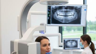
A panoramic X-ray machine is a dental imaging system that captures a broad, curved view of the mouth. It shows the teeth, jawbones, joints, and surrounding structures in one image. During scanning, the X-ray tube and detector rotate around the patient’s head. The patient usually bites a small positioning guide and remains still. This movement creates a panoramic image rather than separate close-up films.

The machine is useful for treatment planning, tooth development checks, implant assessment, and detecting hidden problems. However, it does not replace every dental image. Fine root details may appear less precise than they do in intraoral X-rays.

A poorly positioned patient can produce blurred or distorted results. That limitation is easy to underestimate. Global buyers should review image quality, adjustable exposure settings, patient positioning aids, software compatibility, and local electrical requirements.

Radiation protection, operator training, maintenance access, and documented quality checks also matter.

Tips: Ask for sample images from different patient sizes and clinical cases. Confirm whether the system supports the required image format and workflow. Check service response times before purchase. A lower price may look attractive, but difficult calibration or scarce technical support can increase long-term costs. Always verify compliance with the regulations in the intended market.
Key Features to Compare Before Buying

When comparing the 10 best panoramic X-ray machines for global buyers, image quality deserves more than a high resolution number. Check contrast, distortion control, and anatomical coverage on actual test images. A sharp molar region matters during diagnosis. A wider field of view may help, but it can also increase unnecessary detail. Ask for sample scans from different patient sizes.

Patient positioning should be quick and repeatable. Look for clear bite blocks, forehead supports, adjustable height, and visible alignment guides. Small errors can create blurred arches or uneven exposure. I have found that simple controls often reduce training time. However, overly simplified software may hide useful exposure settings. That trade-off needs honest testing.

Compare exposure control, detector performance, and dose management carefully. Automatic settings should adapt to patient size without removing operator judgment. Review software compatibility, image export formats, network security, and integration with existing systems. Service access is equally important for international buyers. Confirm training, spare-part availability, warranty terms, and response times in writing. A low purchase price can become expensive after installation. Noise, room space, power requirements, and local technical support also affect daily use. Ask operators to test the unit before approval. A brochure rarely shows every practical weakness.

10 Leading Panoramic X-Ray Machines for Global Buyers

10 Leading Panoramic X-Ray Machines for Global Buyers

Choosing among the 10 leading panoramic X-ray machines requires more than comparing prices. Buyers should examine image clarity, patient positioning, exposure control, and daily workflow. A reliable unit should produce consistent dental arches, clear roots, and limited distortion. Modern systems may offer 2D panoramic imaging, cephalometric functions, or optional 3D capabilities. However, advanced features are useful only when staff can operate them confidently. No machine is perfect.

Practical experience shows that installation conditions matter. Check room size, electrical requirements, software compatibility, and shielding guidance before purchase. Look for intuitive controls, adjustable supports, and fast image transfer through accepted medical formats. A strong supplier should provide training, maintenance access, user documentation, and responsive technical support. Buyers should also confirm local registration requirements and service coverage before signing a contract. These details often matter more than attractive specifications.

Tips: Request sample images using different patient sizes. Ask how the machine handles movement and retakes. Compare warranty terms, detector replacement costs, and software updates. A short demonstration can reveal awkward positioning or unclear controls. Independent clinical feedback is valuable, but it may not match every practice. Recheck your priorities after testing the workflow.

10 Best Panoramic X Ray Machines for Global Buyers - 10 Leading Panoramic X-Ray Machines for Global Buyers
No. Representative Configuration Imaging Capability Detector Technology Typical Tube Voltage Typical Tube Current Panoramic Scan Time Optional Cephalometric Arm Typical Field of View Approx. Overall Dimensions Approx. Unit Weight Recommended Use
1 Compact Digital Panoramic Space-saving Standard panoramic, pediatric panoramic, TMJ and bitewing programs Digital CMOS or CCD sensor 60–90 kV 2–16 mA 8–18 seconds Not included Panoramic image area; typically 120–150 mm vertical coverage Approx. 1,000–1,250 W × 1,150–1,450 D × 2,200–2,400 H mm Approx. 65–115 kg General dental clinics with limited installation space
2 General-Purpose Panoramic Everyday diagnosis Panoramic, pediatric, TMJ lateral and frontal, bitewing Direct-conversion CMOS or CCD sensor 60–90 kV 2–16 mA 8–20 seconds Optional on selected configurations Panoramic image area; approximately 130–155 mm vertical coverage Approx. 1,050–1,350 W × 1,200–1,550 D × 2,200–2,500 H mm Approx. 75–140 kg Routine restorative, surgical and orthodontic examinations
3 High-Throughput Panoramic Busy practices Multiple panoramic programs, automatic exposure control, pediatric modes High-resolution CMOS detector 60–90 kV 2–16 mA 8–16 seconds Optional Panoramic image area; typically 130–155 mm vertical coverage Approx. 1,100–1,400 W × 1,250–1,600 D × 2,250–2,500 H mm Approx. 85–150 kg Multi-chair clinics requiring fast patient turnover
4 Panoramic and Cephalometric System Orthodontics Panoramic, lateral cephalometric, optional posteroanterior cephalometric Shared or dual CMOS/CCD detector arrangement 60–90 kV 2–16 mA 8–20 seconds panoramic; 5–15 seconds cephalometric Integrated cephalometric arm Panoramic coverage plus approximately 180–260 mm cephalometric width Approx. 1,600–2,100 W × 1,250–1,650 D × 2,250–2,500 H mm Approx. 125–210 kg Orthodontic departments and dental imaging centers
5 Small-FOV Panoramic and CBCT Implant planning Panoramic, TMJ, endodontic and small-volume 3D CBCT Flat-panel or CMOS-based X-ray detector 60–100 kV 2–16 mA 8–20 seconds panoramic; 5–30 seconds CBCT Not included Typical CBCT volumes: 5 × 5 cm to 8 × 8 cm Approx. 1,100–1,500 W × 1,250–1,650 D × 2,250–2,500 H mm Approx. 120–210 kg Implant, endodontic and localized oral surgery assessment
6 Medium-FOV Panoramic and CBCT Multidisciplinary Panoramic, cephalometric-compatible workflows, dental and maxillofacial CBCT Large-area flat-panel detector 60–110 kV 2–16 mA 8–20 seconds panoramic; 5–40 seconds CBCT Optional on selected configurations Typical CBCT volumes: 8 × 8 cm to 12 × 10 cm Approx. 1,200–1,600 W × 1,300–1,700 D × 2,250–2,550 H mm Approx. 150–260 kg General dentistry, implantology and oral-maxillofacial imaging
7 Low-Dose Pediatric Panoramic Child-friendly Child panoramic, adult panoramic, TMJ and reduced-dose protocols High-sensitivity CMOS or CCD sensor 60–90 kV 1–12 mA 7–18 seconds Not included Panoramic image area; typically 120–150 mm vertical coverage Approx. 950–1,250 W × 1,100–1,450 D × 2,150–2,400 H mm Approx. 60–110 kg Pediatric dentistry and clinics prioritizing dose optimization
8 Orthodontic Panoramic and Cephalometric Growth analysis Panoramic, lateral and posteroanterior cephalometric imaging Dedicated cephalometric sensor with panoramic sensor 60–90 kV 2–16 mA 8–20 seconds panoramic; 5–15 seconds cephalometric Integrated long-arm cephalometric module Panoramic coverage plus approximately 180–300 mm cephalometric width Approx. 1,650–2,200 W × 1,300–1,700 D × 2,250–2,550 H mm Approx. 140–230 kg Orthodontic diagnosis, treatment planning and follow-up
9 Open-Design Panoramic Patient access Standard panoramic, pediatric, TMJ and bitewing programs CMOS or CCD digital detector 60–90 kV 2–16 mA 8–20 seconds Optional Panoramic image area; approximately 125–155 mm vertical coverage Approx. 1,050–1,400 W × 1,200–1,600 D × 2,200–2,500 H mm Approx. 80–150 kg Patients with mobility limitations and high-accessibility requirements
10 Hospital-Grade Panoramic Platform Advanced workflow Panoramic, TMJ, cephalometric-ready workflows and optional CBCT Large-area CMOS or flat-panel detector 60–110 kV 2–16 mA 8–20 seconds panoramic; 5–40 seconds CBCT when equipped Optional integrated module Panoramic coverage; optional CBCT volumes from approximately 5 × 5 cm to 12 × 10 cm Approx. 1,300–2,200 W × 1,350–1,750 D × 2,300–2,600 H mm Approx. 170–300 kg Hospitals, teaching institutions and comprehensive imaging centers
Note: The figures shown are typical published specification ranges for current digital panoramic and panoramic-combination systems. Exact values depend on configuration, regional regulatory requirements, software options and installation conditions. Final purchasing decisions should be based on the official technical documentation, radiation-safety certification and local approval status.

Clinical, Technical, and Compliance Considerations

When comparing panoramic X-ray machines for global buyers, clinical value should lead the decision. A sharp image is not automatically a useful diagnosis. Evaluate mandibular coverage, contrast, motion control, and artifact reduction using real patient workflows. A stable chin rest, clear bite guides, and adjustable positioning lights can reduce retakes. Pediatric protocols matter. So does patient comfort.

Technical review requires more than checking megapixels. Examine the detector type, exposure range, focal spot performance, and image-processing controls. Confirm whether the system supports DICOM export, secure network integration, and serviceable replacement parts. Ask for measured dose data, not only marketing claims. In busy clinics, a short cycle time helps, but rushed positioning can lower image quality. That trade-off deserves testing.

Compliance differs by country and sometimes by region. Buyers should verify medical-device registration, electrical safety documentation, radiation protection requirements, and local acceptance testing. Installation may require room surveys, shielding calculations, and approval from a competent authority. Keep calibration records and quality-assurance logs. Train operators on exposure selection and emergency procedures. Documentation is often overlooked. That is a real weakness.

Independent clinical testing is valuable before purchase. Request phantom images, sample reports, warranty terms, software-update policies, and response times for technical support. A lower purchase price may hide costly downtime or unavailable parts. Even experienced teams can miss this. Use qualified physicists, radiologists, and regulatory specialists when local rules are unclear. A careful comparison protects image quality, staff safety, and long-term operating reliability.

10 Best Panoramic X-Ray Machines for Global Buyers

Clinical, technical, and compliance priorities for international procurement

This chart presents a practical procurement-weighting model rather than manufacturer performance claims. Buyers should verify image quality, patient dose, field of view, installation requirements, software interoperability, and applicable regulatory documentation. Common reference frameworks include IEC 60601-1, IEC 60601-1-3, IEC 60601-2-63, ISO 14971, ISO 13485, and applicable national registration rules.

How to Select the Best Machine for Your Market

Selecting among the 10 best panoramic X-ray machines requires more than comparing image resolution. Your market, patient volume, clinic space, and service access should guide the decision. A compact clinic may need a narrow footprint and simple positioning controls. A busy dental center may value faster scanning, multiple exposure programs, and durable mechanical parts. Check whether the machine supports your local power supply and installation conditions.

Ask suppliers for documented radiation-safety features, software compatibility, warranty terms, and operator training. Requirements differ across countries, so confirm applicable standards with qualified local professionals before purchasing. Inspect sample images from adult, pediatric, and orthodontic cases. A clear molar region matters more than impressive brochure language. I have found that overly complex systems can slow inexperienced staff, although advanced controls may improve consistency after proper training. That trade-off deserves honest review.

Tips: Measure the room, door width, and ceiling height before ordering. Request a demonstration using realistic patient positioning. Compare service response times, replacement-part availability, and software updates. Calculate the full ownership cost, not only the purchase price. Ask whether staff can complete routine scans without repeated retakes. A lower-priced machine may become expensive when training or downtime is overlooked. Keep a written checklist, and let a radiology or dental professional review it before signing.