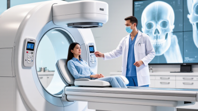
Choosing a 3d dental x ray machine in 2026 requires more than comparing megapixels, scan volume, or promotional prices. The right system must fit your clinical services, patient flow, room dimensions, and staff experience. A busy implant clinic may need large-field CBCT imaging. A general practice may benefit from a compact unit with predictable low-dose protocols. Bigger is not always better.
Look closely at image quality under real conditions. Ask for sample scans showing molars, implant sites, endodontic anatomy, and orthodontic structures. Check whether details remain clear when patients move slightly. Evaluate metal-artifact reduction, voxel size, reconstruction speed, and software usability. Confirm DICOM compatibility before purchase. Your existing sensors, practice-management software, and reporting workflow must communicate reliably. Test the user interface with the people who will operate it daily.
Radiation safety deserves equal attention. Review manufacturer documentation, local compliance requirements, installation needs, and quality-assurance procedures. A reputable supplier should provide training, service records, calibration support, and transparent warranty terms. Speak with experienced dentists who use the same model, not only sales representatives. Their feedback may reveal recurring software delays or difficult maintenance.
There is no perfect machine.
Cost estimates can also mislead. Include installation, shielding assessment, service contracts, software upgrades, and staff training. Consider the machine’s useful life, not merely its purchase price. A practical decision combines diagnostic value, workflow efficiency, patient comfort, and responsible exposure management. Still, every clinic has different priorities. Careful comparison remains necessary, even when the specifications look impressive.
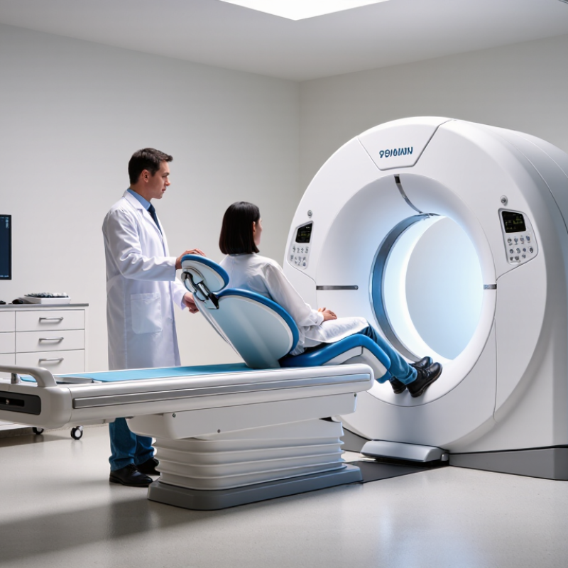
How to Choose a 3D Dental X-Ray Machine in 2026?
Three-dimensional dental X-ray technology uses cone-beam computed tomography, or CBCT, to create volumetric images of teeth, bone, nerves, and airways. Unlike 2D radiographs, CBCT can reveal buccolingual bone width and hidden root anatomy. These details support implant planning, endodontic assessment, oral surgery, and orthodontic diagnosis. The American Association of Endodontists and the American Academy of Oral and Maxillofacial Radiology recommend CBCT only when conventional imaging cannot answer the clinical question.
Dose matters. A review published in Dentomaxillofacial Radiology reported dental CBCT effective doses ranging from about 19 to 1,073 microsieverts, depending on field of view and exposure settings. Smaller fields usually reduce exposure and improve task-specific resolution. Choose adjustable exposure programs, not maximum power. More radiation does not automatically mean better diagnosis.
Look closely at voxel size, field-of-view options, metal-artifact reduction, and reconstruction speed. The European Commission’s Radiation Protection No. 172 guidance emphasizes justification, optimization, and diagnostic quality. Ask whether staff can position a nervous patient comfortably. Check DICOM compatibility and local service support. A beautiful scan is useless if clinicians cannot access it quickly.
Be practical.
Clinical needs should drive specifications, not impressive brochures. A 0.08-millimeter voxel may sound valuable, yet motion can erase that advantage. Training is often underestimated. Review sample scans with a qualified radiologist before purchasing, and compare real patient workflows rather than isolated factory images.
| Evaluation Dimension | Relevant Data or Specification | Clinical Significance | Selection Guidance for 2026 |
|---|---|---|---|
| Imaging technology | Cone-beam computed tomography (CBCT) uses a cone-shaped X-ray beam and a rotating flat-panel detector to create volumetric images. | Provides multiplanar and three-dimensional views of teeth, bone, air spaces, and selected soft-tissue boundaries. | Choose CBCT when three-dimensional information is expected to change diagnosis, treatment planning, or risk assessment. |
| Field of view (FOV) | Common dental CBCT systems offer selectable FOVs from approximately 4 × 4 cm for a limited region to more than 15 × 15 cm for both arches and adjacent structures. | A small FOV generally limits irradiated anatomy and can improve detail for localized tasks; a large FOV covers more anatomy but may increase exposure. | Prefer a system with multiple FOV options rather than using one large volume for every examination. |
| Voxel size | Typical dental CBCT voxel sizes are approximately 0.08–0.40 mm, depending on the selected protocol and device. | Smaller voxels can improve visualization of fine structures, but they may increase image noise, data volume, and dose when higher exposure is used. | Select the smallest practical voxel size for the clinical question, not automatically the smallest available setting. |
| Spatial resolution | High-resolution protocols are designed for fine dental structures; actual performance depends on focal spot, detector, motion, reconstruction, and patient positioning. | Supports assessment of root morphology, narrow canals, periodontal defects, and small osseous structures. | Review measured image-quality data and clinical sample images instead of comparing voxel size alone. |
| Radiation dose | Effective dose varies substantially with FOV, tube voltage, tube current, exposure time, patient size, and reconstruction method. Dental CBCT examinations commonly fall in the broad range of tens to several hundreds of microsieverts. | Dose optimization is essential because CBCT generally exposes the patient to more radiation than a single intraoral or panoramic radiograph. | Compare dose-area product or manufacturer-reported dose data using equivalent FOV and protocol conditions; apply justification and optimization principles. |
| Tube voltage and current | Dental CBCT systems commonly operate within approximately 60–120 kV and use adjustable tube current and exposure time, although available ranges differ by model. | Exposure settings influence penetration, contrast, noise, dose, and suitability for different patient sizes and anatomical regions. | Look for pediatric, adult, and region-specific protocols with accessible exposure controls. |
| Metal artifact management | Modern reconstruction software may include metal-artifact-reduction algorithms, but artifacts from restorations, implants, orthodontic appliances, and surgical hardware cannot be completely eliminated. | Reduced streaking can improve interpretation near metallic objects, while aggressive correction may alter image appearance or obscure detail. | Evaluate raw and corrected images during a clinical demonstration; do not treat artifact reduction as a substitute for correct positioning and protocol selection. |
| Motion control | Typical scan times are commonly in the range of about 5–30 seconds, depending on the system and selected protocol. | Patient movement can produce blur, double contours, and reconstruction artifacts that reduce diagnostic value. | Prioritize stable head supports, clear positioning aids, short acquisition options, and staff training. |
| Implant planning | CBCT can display available bone height and width, cortical boundaries, mandibular canal position, sinus anatomy, and adjacent roots. | Supports implant site evaluation, surgical guide planning, and assessment of anatomical risk. | Select software that supports calibrated measurements, cross-sectional views, implant-planning workflows, and exportable DICOM data. |
| Endodontic applications | High-resolution, limited-FOV scans may assist in evaluating complex canal anatomy, missed canals, resorption, fractures, and persistent periapical disease. | Three-dimensional information can reveal anatomy that is difficult to interpret on two-dimensional images. | Use a limited FOV and appropriate high-detail protocol only when conventional imaging and clinical findings are insufficient. |
| Orthodontic and airway assessment | Large-FOV CBCT can show craniofacial structures, impacted teeth, skeletal relationships, airway spaces, and asymmetry. | Useful for selected orthodontic and maxillofacial questions, but airway volume alone does not establish a sleep-disordered breathing diagnosis. | Choose a large FOV only when the full anatomical region is clinically justified and relevant specialist interpretation is available. |
| Periodontal and osseous evaluation | CBCT may demonstrate buccal and lingual bone plates, furcation regions, fenestrations, dehiscences, and three-wall defects. | Adds information when two-dimensional radiographs do not adequately show the three-dimensional defect or anatomical relationship. | Do not use CBCT as a routine replacement for clinical periodontal examination and conventional radiography. |
| DICOM and software compatibility | DICOM export enables image exchange and integration with compatible planning, surgical-guide, CAD/CAM, and radiology systems. | Interoperability supports referrals, multidisciplinary planning, archiving, and long-term access to patient data. | Confirm unmodified DICOM export, viewer compatibility, data ownership, backup options, and cybersecurity controls before purchase. |
| Workflow and throughput | Workflow depends on scan preparation, patient positioning, acquisition time, reconstruction time, reporting, and image transfer. | Efficient workflow can reduce waiting time and improve consistency without compromising justification or image quality. | Measure the complete patient cycle in a live demonstration rather than evaluating scan time alone. |
| Installation requirements | Requirements may include room dimensions, structural support, electrical supply, radiation shielding assessment, network access, and regulatory approval. | Insufficient space, power, shielding, or connectivity can delay installation and increase operating costs. | Obtain a site survey and verify local radiation-safety, electrical, building, and medical-device requirements before signing an order. |
| Quality assurance and maintenance | Routine quality-control checks should monitor image quality, calibration, geometric accuracy, exposure consistency, and software performance. | Regular testing helps identify drift, repeat scans, artifacts, and safety issues at an early stage. | Compare service response times, preventive-maintenance plans, calibration procedures, training, warranty coverage, and total cost of ownership. |
| Decision rule | The appropriate system is determined by clinical indications, required FOV, image quality, dose optimization, workflow, compatibility, compliance, and budget. | No single specification guarantees the best clinical result for every examination. | Build a protocol-based evaluation using representative cases, objective dose and image-quality data, staff usability, and lifecycle cost. |
Note: Technical ranges are representative industry values and can vary by system, protocol, patient size, reconstruction method, and local regulations. Clinical use should follow applicable radiation-protection guidance and professional judgment.