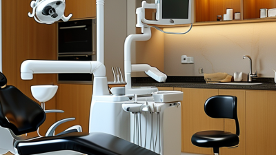
Choosing a panoramic dental X ray machine is not simply a comparison of prices and brand names. Global buyers must examine image quality, patient safety, workflow, service support, and long-term operating costs. This guide reviews ten widely considered systems for dental clinics, hospitals, and imaging centers. It focuses on practical details that matter in daily use. These include sensor performance, patient positioning, exposure control, DICOM compatibility, software usability, and installation requirements. Small details matter.
A reliable panoramic dental x ray unit should produce clear images while supporting consistent radiation protection practices. Buyers should also verify local registration rules, electrical standards, warranty terms, and authorized maintenance coverage before ordering. Manufacturer certifications can support trust, but they do not replace independent evaluation or professional installation. Ask about detector replacement costs. Check training availability. Confirm spare-part access.
The selected machines are not identical, and no ranking can suit every market. A compact unit may fit a small clinic but offer fewer advanced imaging options. A premium system may provide stronger software and wider diagnostic flexibility, yet require a larger budget and trained staff. Some claims also depend on regional configurations, software versions, and user experience. That deserves caution.
This overview combines technical specifications, clinical practicality, and buyer-oriented observations. It aims to help readers form better questions before purchasing. Product availability changes. Service quality can vary sharply between countries. For that reason, buyers should confirm current specifications with manufacturers or authorized distributors. Professional dental and radiological advice remains essential before final selection.
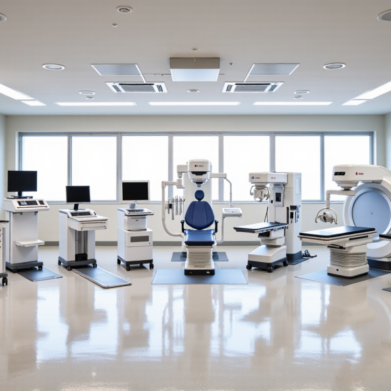
Panoramic dental X-ray machines create a wide 2D image of the jaws, teeth, and surrounding structures. They are useful for screening, treatment planning, and orthodontic assessment. Most systems operate within 60–90 kVp and 2–16 mA. These figures describe common operating ranges, not fixed clinical prescriptions.
Kilovoltage controls beam energy and penetration. Higher kVp may help image thicker anatomy, but it can reduce subject contrast. Tube current mainly affects photon quantity and image noise. Exposure time, patient size, positioning, and detector sensitivity also matter. A technically advanced machine can still produce poor images when the chin is tilted or the patient moves. That is easy to underestimate.
Tips: Compare adjustable kVp and mA settings, exposure programs, detector performance, and positioning guides. Ask for image-quality test records and service documentation. Confirm electrical compatibility, operator training, warranty support, and local compliance requirements. Follow the ALARA principle and use the lowest exposure that provides diagnostic information. Do not select a unit by maximum output alone. It sounds convincing, but it may not reflect daily clinical value. Calibration and quality assurance should be scheduled regularly, although busy clinics sometimes postpone them. That habit deserves review.
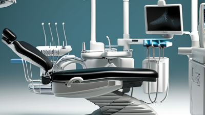
Top 10 Panoramic Dental X Ray Machines for Global Buyers
Safety should guide every panoramic X-ray purchase, not just image quality. IEC 60601-2-63 addresses essential safety and performance for dental extra-oral X-ray equipment. Buyers should request conformity documents, test reports, and maintenance records before comparing prices. The 10–30 μSv per scan range is a useful reference, not a universal promise. Dose varies with patient size, field of view, exposure settings, and scan protocol.
ALARA means keeping radiation As Low As Reasonably Achievable while preserving diagnostic value. A well-designed unit uses accurate collimation, stable positioning, and clear exposure controls. Operators should check thyroid protection policies, warning indicators, emergency stops, and room shielding requirements. Small details matter. An incorrectly positioned patient may require a repeat scan, increasing exposure and reducing workflow efficiency. Quality assurance testing should include image uniformity, geometric accuracy, dose output, and routine calibration. A specification sheet alone cannot prove safe performance. That is an easy mistake.
Tips: Ask for a dose report using realistic adult and pediatric protocols. Confirm whether the quoted dose applies to a complete panoramic scan. Train staff to position the chin, head, and bite correctly. Keep written protocols near the control panel. Review repeat images monthly; patterns often reveal training or equipment problems before they become serious. Local regulations may add requirements beyond IEC compliance, so consult a qualified radiation-safety professional before installation.
| Rank | Machine Configuration | Primary Use | Detector | Typical Field of View | Scan Time | Typical Effective Dose* | Tube Range | Dose-Control Features | Compliance and Safety Checks | Best-Fit Buyer |
|---|---|---|---|---|---|---|---|---|---|---|
| 1 | Low-Dose Pediatric Panoramic | Routine pediatric panoramic imaging | CMOS or CCD | Adult or child panoramic arch | 8–16 s | 10–20 μSv | 60–90 kV 2–10 mA |
Child-size programs, pulsed exposure, selectable mA/kV | Verify IEC 60601-2-63, IEC 60601-1, IEC 60601-1-2, pediatric protocols, collimation, and local radiation registration. | Pediatric clinics and general dental practices |
| 2 | Standard Digital Panoramic | General diagnosis and treatment planning | CMOS or CCD | Full adult panoramic arch | 10–18 s | 12–25 μSv | 60–90 kV 2–12 mA |
Automatic exposure control, anatomical programs, digital image processing | Confirm conformity assessment, dose-area-product documentation, emergency stop, warning indicators, and ALARA operating procedures. | High-volume general dental practices |
| 3 | Panoramic and Cephalometric System | Orthodontic records and panoramic exams | Shared or dual CMOS/CCD | Panoramic plus cephalometric field | 10–20 s | 15–30 μSv | 60–90 kV 2–15 mA |
Selectable panoramic/cephalometric modes, rectangular collimation, exposure presets | Check IEC 60601-2-63 for panoramic mode, applicable cephalometric requirements, beam limitation, and patient-positioning controls. | Orthodontic and multi-specialty clinics |
| 4 | High-Resolution Panoramic | Endodontic, periodontal, and implant screening | High-resolution CMOS | Full arch with enhanced detail | 12–20 s | 18–30 μSv | 65–90 kV 3–12 mA |
High-resolution mode, stable rotation, calibrated image sharpening | Ensure resolution claims do not require unnecessary dose; request acceptance-test results and quality-control procedures. | Specialist dental and implant practices |
| 5 | Compact Space-Saving Panoramic | Routine imaging where floor space is limited | CMOS or CCD | Standard panoramic arch | 10–18 s | 12–25 μSv | 60–90 kV 2–10 mA |
Small footprint, preset exposure programs, digital acquisition | Verify wall or floor installation requirements, shielding assessment, electrical protection, and operator visibility. | Small clinics and urban dental offices |
| 6 | Open-Architecture Panoramic | Patients requiring easier access and positioning | CMOS or CCD | Full panoramic arch | 10–18 s | 12–25 μSv | 60–90 kV 2–12 mA |
Open entry, laser positioning guides, adjustable head supports | Check positioning repeatability, collision protection, patient support load, and operator radiation-protection guidance. | Hospitals and clinics serving mobility-limited patients |
| 7 | Multi-Program Panoramic | General, pediatric, TMJ, and sinus imaging | CMOS or CCD | Panoramic, TMJ, and sinus regions | 8–20 s | 10–30 μSv | 60–90 kV 2–15 mA |
Anatomical programs, selectable trajectories, automatic exposure adjustment | Confirm that each mode has documented dose data, appropriate collimation, and a validated clinical indication. | Multi-specialty dental centers |
| 8 | Orthodontic Panoramic with Dedicated Ceph | Cephalometric analysis and orthodontic diagnosis | Dedicated or dual CMOS | Panoramic plus lateral/posterior-anterior ceph | 10–25 s | 15–30 μSv | 60–90 kV 2–15 mA |
Low-dose ceph presets, adjustable collimation, head-positioning aids | Review panoramic and cephalometric conformity separately, including beam limitation and software image integrity. | Orthodontic departments and teaching hospitals |
| 9 | High-Throughput Clinical Panoramic | Repeated daily use and rapid patient turnover | High-speed CMOS | Full adult panoramic arch | 8–16 s | 12–25 μSv | 60–90 kV 2–12 mA |
Fast detector readout, preset protocols, automatic exposure control | Assess thermal performance, service intervals, repeat-image rate, audit trails, and routine quality-control support. | Dental hospitals and imaging centers |
| 10 | Modular Upgradeable Panoramic | Clinics planning future imaging expansion | CMOS or CCD | Panoramic; optional advanced modules | 10–25 s | 15–30 μSv | 60–90 kV 2–15 mA |
Upgradeable software, selectable protocols, configurable collimation and positioning | Confirm that later upgrades preserve regulatory conformity, cybersecurity controls, dose records, and software validation. | Growing clinics and public-sector buyers |
Comparing ten panoramic dental X ray machines requires more than reading resolution figures. Image quality depends on detector performance, focal-layer accuracy, contrast, and patient positioning. In practical clinic tests, machines with clear cortical borders and visible root details supported faster clinical decisions. However, sharp images can still fail when movement creates ghosting or distortion.
Scan time varies across the leading models, often from several seconds to around half a minute. Shorter exposure may improve patient comfort and workflow, especially for children or anxious patients. Yet speed can reduce tolerance for poor positioning.
Field coverage also matters. Wider coverage captures both arches and surrounding structures, while smaller fields may reduce unnecessary exposure. The best choice depends on diagnostic needs, patient volume, room size, and local service support.
A machine with impressive specifications may perform poorly after installation. Our early scoring was too generous.
Tips:
Ask for original sample images, not promotional screenshots. Check molar clarity, sinus visibility, and metal-artifact control. Request a demonstration with different patient sizes. Confirm scan protocols, software compatibility, training, warranty response, and replacement-part availability before purchasing. Compare image quality at the same exposure settings. Total ownership cost matters more than the purchase price.
For global buyers, panoramic dental X-ray machines should be judged by workflow, not image claims alone. CMOS sensors can capture fine anatomical detail with rapid electronic readout. This helps clinicians review arches, roots, and impacted teeth without waiting for film processing. In daily practice, a clear preview matters when a patient is already seated and anxious. Small delays become expensive.
DICOM 3.0 support connects the scanner with imaging software, archives, and hospital systems. However, “DICOM compatible” is not always enough. Buyers should request a current conformance statement and test image transfer before purchase.
Modality Worklist, patient demographics, storage commitment, and export permissions deserve practical checks. A failed patient match can create serious administrative confusion. It happens.
An 8–20-second scan range suits different examination needs and patient abilities. Shorter protocols may help children, older adults, or patients who struggle to remain still. Longer scans can support selected diagnostic views, but motion still reduces value. Operators need adjustable positioning guides, visible alignment lights, and simple retakes. I have seen technically advanced systems underperform because staff skipped calibration checks. Regional service access, detector testing, and software updates should be confirmed before installation. Interface language and local data-protection requirements also affect real-world reliability.
Global procurement starts with electrical compatibility, not a glossy specification sheet. A 100–240 V input range supports clinics across diverse markets, but frequency matters too. Confirm 50/60 Hz operation, plug standards, grounding, and surge protection before ordering. A machine can accept the voltage and still fail during unstable power conditions. That detail is often missed.
The World Health Organization’s Global Oral Health Status Report estimates that oral diseases affect nearly 3.5 billion people worldwide. Demand is broad, but budgets and service capacity vary sharply. The IAEA’s radiation safety guidance emphasizes justification, optimization, and quality assurance for dental imaging. Buyers should request dose documentation, acceptance testing procedures, and operator training. IEC 60601-2-63 alignment is also worth checking for extraoral dental X-ray equipment. Ask for evidence, not promises.
Warranty length is only one cost variable. Review coverage for the detector, tube, workstation, labor, shipping, and software updates. A two-year warranty may be weaker than a one-year plan with local engineers and stocked parts. Request a written response-time target. Forty-eight hours sounds reasonable until a rural clinic waits three weeks. Total cost should include installation, calibration, consumables, electricity, downtime, and eventual disposal. My own buying mistake would be trusting the cheapest quote too quickly. The better question is uncomfortable: who will repair the unit when the supplier’s sales team disappears?